Activation Postganglionic Sympathetic Fibres
Autonomic nervous system: see nervous system,network of specialized tissue that controls actions and reactions of the body and its adjustment to the environment. Virtually all members of the animal kingdom have at least a rudimentary nervous system. Click the link for more information. Autonomic nervous systemThe part of the nervous system that innervates smooth and cardiac muscle and the glands, and regulates visceral processes including those associated with cardiovascular activity, digestion, metabolism, and thermoregulation. The autonomic nervous system functions primarily at a subconscious level. It is traditionally partitioned into the sympathetic system and the parasympathetic system, based on the region of the brain or spinal cord in which the autonomic nerves have their origin. The sympathetic system is defined by the autonomic fibers that exit thoracic and lumbar segments of the spinal cord.
The parasympathetic system is defined by the autonomic fibers that either exit the brainstem via the cranial nerves or exit the sacral segments of the spinal cord. See,The defining features of the autonomic nervous system were initially limited to motor fibers innervating glands and smooth and cardiac muscle. This definition limited the autonomic nervous system to visceral efferent fibers and excluded the sensory fibers that accompany most visceral motor fibers. Although the definition is often expanded to include both peripheral and central structures (such as the hypothalamus), contemporary literature continues to define the autonomic nervous system solely as a motor system.
However, from a functional perspective, the autonomic nervous system includes afferent pathways conveying information regarding the visceral organs and the brain areas (such as the medulla and the hypothalamus) that interpret the afferent feedback and exert control over the motor output back to the visceral organs. See Autonomic Nervous System.
(also vegetative nervous system), the part of the nervous system that regulates the organs of blood circulation, respiration, digestion, excretion, and reproduction, as well as metabolism, thereby regulating the functional state of all the tissues of vertebrate animals and man.The term “vegetative nervous system” was introduced by the French biologist M. Bichat (1800), who divided the nervous system into animal (somatic), that is, regulating the functions peculiar to animals alone and responsible for sensations and body movements, and vegetative, regulating the main vital processes—nutrition, respiration, reproduction, and growth (peculiar not only to animals but to plants as well). The functions regulated by the vegetative nervous system may not be performed or halted voluntarily. Hence the English physiologist J. Langley called it autonomic.
However, the “autonomy” of the autonomic nervous system with respect to the higher divisions of the brain is extremely relative because impulses traveling from the cortex of the cerebral hemispheres to the centers of the autonomic nervous system may alter the functioning of the internal organs. Each complex reaction of the organism, any behavioral act, voluntary or involuntary, includes the sensing of stimuli, sensations, body movements, and the functional changes in the organs innervated by the autonomic nervous system.The autonomic nervous system is divided into two parts according to anatomical and physiological features: sympathetic and parasympathetic. The centers of the sympathetic nervous system (SNS) are located in the thoracic and lumbar segments of the spinal cord. The centers of the parasympathetic nervous system (PNS) are located in the midbrain and medulla oblongata and in the sacral segments of the spinal cord. The main nerve of the PNS—the one that transmits the influence of the PNS to many organs of the body—is the vagus nerve. The sympathetic and parasympathetic centers are subordinated to the centers of the autonomic nervous system located in the diencephalon—in the hypothalamus, which coordinates the functions of both parts of the autonomic nervous system and regulates metabolism and the functions of many organs and systems. The highest control over the autonomic nervous system is exerted by the centers of the cerebral hemispheres that ensure the integrated reaction of the body and maintain through the autonomic nervous system the necessary correspondence of intensity between the vital processes—metabolism, blood circulation, respiration, and so on—and the requirements of the body.All the sympathetic and parasympathetic nervous pathways to the periphery are formed by two successively connected nerve cells (neurons).
The cell body of the first neuron is in the midbrain, medulla oblongata, or spinal cord. The long process (axon) of the first neuron ends in nerve cells on the periphery that form ganglia.

Within the ganglion is the cell body of the second neuron, whose process transmits impulses to the organ it innervates. (The fibers of the first neuron are called preganglionic; the fibers of the second are called postganglionic.) Thus, the nerves of the autonomic nervous system, unlike the motor nerves of the striated muscles which are continuous after emerging from the central nervous system, have a break in their fibers. The peripheral neurons of the SNS form ganglia on both sides of the spinal cord (sympathetic trunks) and in the neck and abdominal cavity. The peripheral neurons of the PNS are located directly in the organs they innervate. Every preganglionic fiber ends in many neurons in the ganglia, which greatly widens the zone of influence of the preganglionic neurons.
Every postganglionic neuron has endings formed by various processes of preganglionic neurons. Hence the impulses reaching the nerve cell via different nerve fibers may be summated.The preganglionic nerve fibers have a thin medullated, or myelin, sheath and are 2-3½microns in diameter—that is, they are far thinner than the motor fibers innervating the striated muscles.
Most of the postganglionic fibers lack a myelin sheath and are even thinner. The nerve fibers of the autonomic nervous system are characterized by low excitability and a low rate of conduction of excitation. The endings of the parasympathetic and sympathetic fibers differ with respect to the chemical transmitters of nervous impulses (mediators) formed in them.
Activation Postganglionic Sympathetic Fibers Of The Heart
The mediator acetylcholine is formed in the endings of all the parasympathetic nerve fibers and the preganglionic sympathetic nerve fibers, as well as the postganglionic sympathetic fibers innervating the sweat glands. The mediator norepinephrine is formed in the endings of the postganglionic sympathetic fibers, except those innervating the sweat glands. The English physiologist H. Dale suggested dividing the nerve fibers into cholinergic and adrenergic ones, depending on the chemical nature of the mediators formed in their endings. After transection and degeneration of sympathetic or parasympathetic nerves, the sensitivity of the denervated organs to the corresponding mediators increases sharply.

The organ deprived of sympathetic innervation is particularly sensitive to norepinephrine and epinephrine, while the organ deprived of parasympathetic innervation is particularly sensitive to acetylcholine.Excitation of the SNS stimulates the body; excitation of the PNS helps to restore the resources used up by the body. The SNS and PNS are functional antagonists and therefore exert opposite influences on many organs.
Thus, under the influence of impulses traveling along the sympathetic nerves, heart contractions accelerate and intensify, blood pressure in the arteries rises, glycogen is broken down in the liver and muscles, blood glucose increases, the pupils dilate, sensitivity of the sensory organs and efficiency of the central nervous system increase, the bronchi constrict, stomach and intestinal contractions are inhibited, the secretion of gastric and pancreatic juices diminishes, and the urinary bladder relaxes and evacuation is inhibited. Under the influence of impulses arriving via the parasympathetic nerves, heart contractions slow and weaken, arterial pressure drops, blood glucose decreases, stomach and intestinal contractions are stimulated, the secretion of gastric and pancreatic juices is intensified, and so on. The activity and state of certain organs are controlled solely by the sympathetic nerves—for example, the sweat glands, most blood vessels (except those of the tongue, salivary glands, and genitalia, the blood vessels of which are constricted by sympathetic nerves and dilated by parasympathetic nerves), adrenal glands, and uterus.The autonomic nervous system has a threefold effect on organs: activating, correcting, and adaptotrophic. The activating effect is manifested by impulses stimulating the activity of an organ that functions periodically (for example, stimulation of secretion by the sweat glands under the influence of sympathetic nerves). The correcting effect is manifested by an intensification or weakening of the activity and state of excitation (tonus) of organs that possess automatism and that function continuously or are in a constant state of excitation (for example, the effect of the autonomic nervous system on heart action and the state of the blood vessels). The adaptotrophic function of the autonomic nervous system, mainly the SNS, consists of regulating metabolism and the functional state (excitability, efficiency) of organs and tissues; it prepares the body for activity and adjusts the work of the organs to external conditions and current needs of the body.The role of the SNS in adapting the body to various situations requiring physical exertion was demonstrated in experiments on animals from whom both sympathetic trunks and all the sympathetic ganglia were removed (sympathectomy). While resting, such animals scarcely differ from normal ones.

But during vigorous muscular exertion, overheating, chilling, loss of blood, or emotional stress, animals whose organs have been deprived of sympathetic influences have little endurance. Because of impairment of thermoregulation, they tolerate abrupt fluctuations in external temperature less easily than do normal animals. They become chilled sooner when exposed to cold and become overheated sooner when exposed to heat.The autonomic nervous system (mainly its sympathetic division) becomes excited in extreme, life-threatening situations that require maximum exertion of the body’s forces—for example, suffocation, loss of blood, attack by an enemy, and trauma:—and in emotional reactions. This accounts for the acceleration and intensification of heart contractions, dilatation of the skin blood vessels, and reddening of the face in joy; for the pallor, sweating, gooseflesh, inhibition of gastric secretion, and change in intestinal peristalsis in fear; for pupil dilatation in anger, pain; and so on.The physiological manifestations of emotions are caused mainly by excitation of the SNS. For example, in emotional stress and excitation of the central nervous system provoked by pain, impulses reaching certain endocrine glands along the fibers of the autonomic nervous system intensify the secretion of hormones into the blood.
The American physiologist W. Cannon showed that emotional reactions increase the entry into the blood of epinephrine from the adrenal glands under the influence of impulses reaching them from the sympathetic nerves. Excitation of the autonomic centers of the hypothalamus resulting from pain stimulates the entry of various hormones from the pituitary, thyroid, and other glands into the blood. Release into the blood of epinephine (which affects many organs like the sympathetic nerves), vasopressin (which constricts the blood vessels and halts urination), and other hormones under the influence of the autonomic nervous system supplements and intensifies its direct action on the functions of various organs stimulated by the entry of nerve impulses. This is how neurohormonal regulation of the body’s activity is effected. Thus, the activity of the autonomic nervous system consists of the complementary interaction of its sympathetic and parasympathetic divisions.
The SNS largely stimulates the processes associated with the release of energy (dissimilation), with activity, whereas the PNS stimulates the processes associated with the accumulation of energy and matter (assimilation).
Key Points. Postganglionic fibers in the sympathetic division are adrenergic and use norepinephrine (also called noradrenalin) as a neurotransmitter. Key Terms.
postganglionic fiber: In the autonomic nervous system, these are the fibers that run from the ganglion to the effector organ. cholinergic: Pertaining to, activated by, producing, or having the same function as acetylcholine. adrenergic: Containing or releasing adrenaline. postganglionic neuron: A nerve cell that is located distal or posterior to a ganglion.In the autonomic nervous system, fibers from the ganglion to the effector organ are called postganglionic fibers. The post-ganglionic neurons are directly responsible for changes in the activity of the target organ via biochemical modulation and neurotransmitter release.The neurotransmitters used by postganglionic fibers differ.
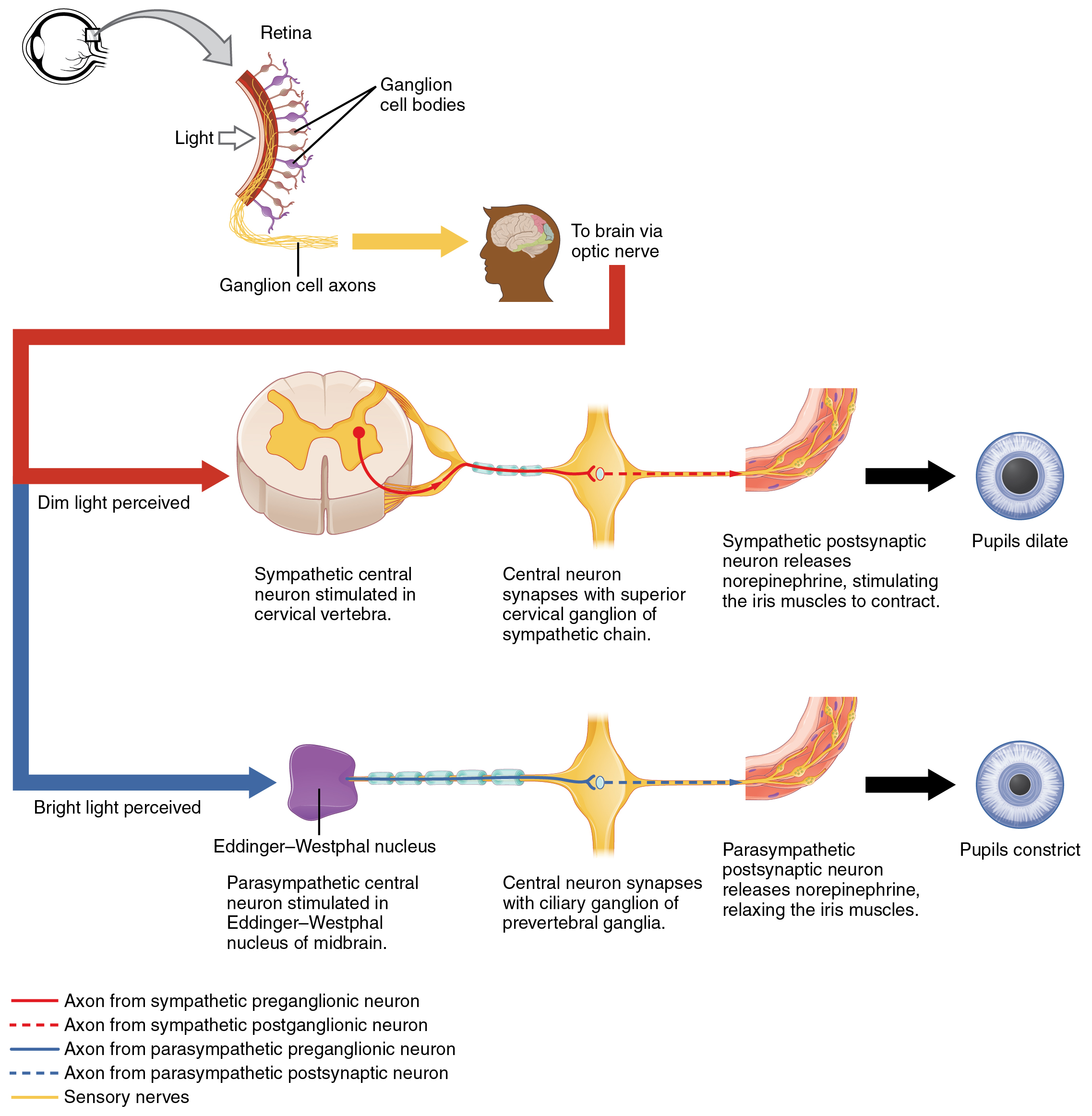
In the parasympathetic division, they are cholinergic and use acetylcholine as their neurotransmitter. In the sympathetic division, most are adrenergic, meaning they use norepinephrine as their neurotransmitter.Postganglionic nerve fibers: In the autonomic nervous system, preganglionic fibers (shown in light blue) carry information from the CNS to the ganglion. The Sympathetic FibersAt the synapses within the ganglia, the preganglionic neurons release acetylcholine, a neurotransmitter that activates nicotinic acetylcholine receptors on postganglionic neurons. In response to this stimulus, postganglionic neurons—with two important exceptions—release norepinephrine, which activates adrenergic receptors on the peripheral target tissues. The activation of target tissue receptors causes the effects associated with the sympathetic system.The two exceptions mentioned above are the postganglionic neurons of sweat glands and the chromaffin cells of the adrenal medulla. The postganglionic neurons of sweat glands release acetylcholine for the activation of muscarinic receptors.
The chromaffin cells of the adrenal medulla are analogous to post-ganglionic neurons—the adrenal medulla develops in tandem with the sympathetic nervous system and acts as a modified sympathetic ganglion. Within this endocrine gland, the pre-ganglionic neurons create synapses with chromaffin cells and stimulate the chromaffin cells to release norepinephrine and epinephrine directly into the blood.Presynaptic nerves’ axons terminate in either the paravertebral ganglia or prevertebral ganglia. In all cases, the axon enters the paravertebral ganglion at the level of its originating spinal nerve.After this, it can then either create a synapse in this ganglion, ascend to a more superior ganglion, or descend to a more inferior paravertebral ganglion and make a synapse there, or it can descend to a prevertebral ganglion and create a synapse there with the postsynaptic cell. The postsynaptic cell then goes on to innervate the targeted end effector (i.e., gland, smooth muscle, etc.).Because paravertebral and prevertebral ganglia are relatively close to the spinal cord, presynaptic neurons are generally much shorter than their postsynaptic counterparts, which must extend throughout the body to reach their destinations.
The Parasympathetic FibersThe axons of presynaptic parasympathetic neurons are usually long. They extend from the CNS into a ganglion that is either very close to or embedded in their target organ. The LibreTexts libraries are and are supported by the Department of Education Open Textbook Pilot Project, the UC Davis Office of the Provost, the UC Davis Library, the California State University Affordable Learning Solutions Program, and Merlot. We also acknowledge previous National Science Foundation support under grant numbers 1246120, 1525057, and 1413739.
Unless otherwise noted, LibreTexts content is licensed. Have questions or comments? For more information contact us at or check out our status page at.